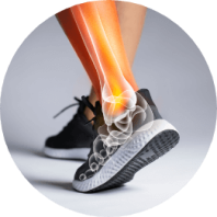
Midfoot
Fractures
- Home
- Conditions We Treat
- Foot & Ankle
- Midfoot Fractures
What are midfoot fractures?
A midfoot fracture involves injury to the bones or ligaments in the central part of the foot, most often at the Lisfranc joint complex. This joint is crucial for stabilising the foot arch and transferring body weight during standing and walking. Injuries may include fractures of the metatarsals, cuneiforms or cuboid bones, as well as dislocations or ligament tears.
Although uncommon, midfoot fractures are clinically significant because they are frequently mistaken for simple sprains. Missed or delayed diagnosis can lead to persistent pain, progressive deformity and arthritis, all of which affect mobility and quality of life. Early recognition and appropriate treatment are therefore essential for good long-term outcomes.
Understanding the anatomy of the midfoot is key to recognising how these injuries occur and why they can be so disabling.
Anatomy of the midfoot
The midfoot is formed by the three cuneiform bones and the cuboid, which articulate with the five metatarsals at the Lisfranc joint. This joint is divided into three functional columns:
- Medial column — the first metatarsal and medial cuneiform.
- Central column — the second and third metatarsals with the intermediate and lateral cuneiforms.
- Lateral column — the fourth and fifth metatarsals with the cuboid.
A complex arrangement of tarsometatarsal, intermetatarsal and intertarsal ligaments provides stability to these articulations, while allowing controlled movement. Together, these structures maintain the integrity of the foot arch and bear the body’s weight during gait.
Types of midfoot fractures
Midfoot fractures, especially those involving the Lisfranc joint, may present in several patterns depending on the direction and force of injury:
- Avulsion fractures — a small fragment of bone is pulled away where a ligament attaches, usually due to twisting or indirect force.
- Shear (split) fractures — the bone splits when opposing forces act on its surfaces. These may occur in the coronal (front-to-back) or sagittal (side-to-side) plane.
- Impaction fractures — the bone ends are compressed or “driven into” one another. This may involve one joint (uniarticular) or extend across multiple joints (biarticular) in high-energy injuries.
These patterns can involve several bones of the midfoot, including the cuboid, cuneiforms and navicular. Injuries often affect multiple bones at once. The medial cuneiform and the base of the second metatarsal are particularly vulnerable, as they form the central stabilising point of the Lisfranc joint.
Clinical classification systems
In clinical practice, surgeons often use the Hardcastle and Myerson classification to describe Lisfranc injuries more precisely:
- Type A — total incongruity, where all the metatarsals are displaced in the same direction.
- Type B — partial incongruity, where only some metatarsals are displaced.
- Type C — divergent, where the metatarsals are displaced in different directions.
This system helps guide treatment decisions and prognosis, especially in complex or high-energy injuries.

What causes midfoot fractures?
Midfoot fractures, particularly those involving the Lisfranc joint, can result from a variety of injury mechanisms. They are broadly divided into high-energy trauma and low-energy twisting injuries:
- High-energy trauma — these injuries often occur during motor vehicle accidents, falls from height or industrial crush accidents. The force is transmitted directly to the midfoot, causing complex fractures, dislocations or both. Because of the severity of the impact, high-energy injuries are frequently associated with extensive soft tissue damage and carry a poorer prognosis.
- Low-energy or indirect trauma — more subtle injuries may occur when the foot is planted while the body twists, such as during sports like football, rugby or gymnastics. The sudden rotational stress disrupts the tarsometatarsal ligaments and may cause fractures of the metatarsal bases. These injuries are sometimes mistaken for simple sprains, leading to delayed diagnosis.
- Direct trauma — a heavy object falling onto the foot or the foot being run over or crushed, can directly fracture the midfoot bones. This mechanism typically produces severe comminuted fractures and carries a higher risk of long-term complications.
Both direct and indirect mechanisms are important, but indirect twisting injuries are more common, whereas direct high-energy crush injuries tend to result in more severe deformity and disability if not managed promptly.
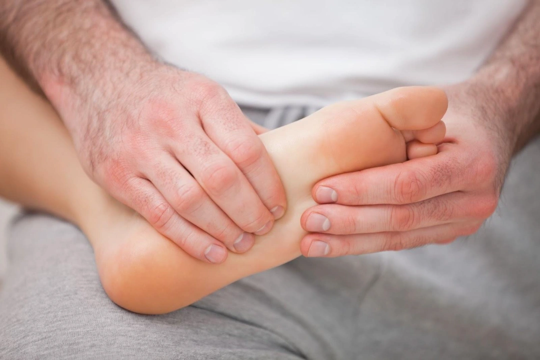
What are the symptoms of a midfoot fracture?
Midfoot fractures, especially Lisfranc injuries, often present with symptoms that resemble a simple sprain. This similarity is one of the main reasons they are frequently overlooked or diagnosed late. Typical symptoms include:
- Pain — usually felt in the middle of the foot, often worsening with weight-bearing or when pushing off the foot.
- Swelling — rapid swelling across the midfoot, sometimes extending to the ankle.
- Bruising — bruising on the top and particularly on the sole of the foot is a strong indicator of a Lisfranc injury.
- Difficulty walking — many patients cannot put weight on the affected foot or find it extremely painful to do so.
- Foot deformity — in severe injuries, the arch of the foot may collapse, or the midfoot may appear widened or misaligned.
Red flag signs — when to seek urgent medical attention
- Inability to bear any weight on the injured foot.
- Bruising on the sole of the foot (a classic sign of Lisfranc injury).
- Noticeable deformity or collapse of the foot arch.
- Severe pain that does not improve with rest and elevation.
Because untreated midfoot fractures can result in chronic pain, arthritis, and long-term disability, it is crucial to seek prompt medical assessment if these warning signs are present.
Who is at risk of midfoot fractures in Singapore?
While midfoot fractures can happen to anyone after sufficient trauma, certain groups face a higher risk because of either their activities or underlying bone health:
- Motorcyclists and road users — high-energy accidents, particularly involving motorcycles or cars, commonly cause midfoot and Lisfranc injuries due to direct impact or crush forces.
- Athletes — sports that involve sudden twisting, pivoting or falls (e.g. football, rugby, gymnastics, equestrian sports) increase the risk of ligamentous and bony midfoot injuries.
- High-risk occupations — jobs in construction, manufacturing or heavy industry expose workers to crush injuries from falling objects or machinery accidents.
- People with reduced bone strength — individuals with osteoporosis, prior fractures, chronic steroid use or post-menopausal bone loss are more vulnerable. In these cases, even relatively minor trauma, such as tripping or twisting the foot, can cause a fracture.
Both high-energy trauma and compromised bone health raise the likelihood of midfoot fractures. Recognising these risks is important, as early assessment and treatment significantly improve long-term outcomes.
What are the complications of midfoot fractures?
If midfoot fractures are missed or not treated properly, they can lead to long-term complications that affect mobility and quality of life:
- Post-traumatic arthritis — the most common complication. Damage to the cartilage and joint surfaces during injury, especially if left untreated or inadequately reduced, often leads to early arthritis and persistent stiffness.
- Chronic pain — ongoing pain may develop from arthritis, malalignment or residual instability. This can interfere with daily activities and reduce overall quality of life.
- Malunion and nonunion — malunion occurs when bones heal in the wrong position, while nonunion means the fracture fails to heal altogether. Both can cause pain, instability and difficulty bearing weight on the foot.
- Acquired flatfoot (planovalgus deformity) — if the midfoot arch collapses after injury, patients may develop a flatfoot deformity. This can lead to altered walking patterns, difficulty with footwear and pain during prolonged standing or walking.
Delayed diagnosis or treatment significantly increases the risk of these complications. Prompt medical evaluation and appropriate management are essential to preserve foot function and prevent long-term disability.
How are midfoot fractures diagnosed?
Midfoot fractures are often difficult to detect because their symptoms resemble simple sprains. A prompt and accurate diagnosis by a medical professional is essential, as missed injuries can lead to chronic pain, deformity, and arthritis. Diagnosis usually involves a combination of clinical assessment and imaging tests:
- Physical examination — the doctor will check for swelling, bruising (especially on the sole of the foot, a classic sign of Lisfranc injury) and localised tenderness over the midfoot. Stressing the forefoot or asking the patient to bear weight may reproduce pain. In severe cases, misalignment or collapse of the arch may be visible.
- X-ray (radiograph) — standard weight-bearing X-rays are taken in anteroposterior, lateral and 30° oblique views. These allow the doctor to identify fractures, subtle joint widening or displacement of the metatarsal bases. Non–weight-bearing films may miss injuries.
- CT scan — CT is more sensitive than X-ray and provides detailed information on small fractures, displacement and fracture orientation. It is especially useful in high-energy injuries and for planning surgery.
- MRI scan — MRI is reserved for cases where ligamentous injury is suspected or when X-ray/CT results are inconclusive. It is the best test for assessing soft tissue and cartilage damage.
If you have persistent midfoot pain after an injury and initial X-rays appear normal, ask your doctor whether advanced imaging such as a CT or MRI scan is needed. This can help avoid a missed diagnosis and long-term complications.
What are the treatment options for midfoot fractures in Singapore?
Treatment depends on the severity and stability of the injury. Stable, nondisplaced fractures may heal with conservative management, while unstable or displaced fractures usually require surgery to restore alignment and prevent long-term complications.
Non-surgical treatment — when the injury is stable
Non-surgical treatment is considered when the midfoot fracture is stable and properly aligned, meaning the bones have not shifted out of place. This usually applies to nondisplaced fractures, stress-related fractures, or low-energy injuries that do not disrupt the midfoot joints. In such cases, the injury can often heal successfully with immobilisation and careful monitoring rather than surgery.
- Immobilisation — immobilisation keeps the foot protected while the fracture heals naturally. A below-knee cast or walker boot is applied with strict non-weight-bearing for around 6 weeks, followed by protected weight-bearing in a boot for another 4–6 weeks.
- Rehabilitation — rehabilitation ensures that stiffness is minimised and function is restored. Early ankle and toe movements may be introduced, progressing to strengthening and gait training once partial weight-bearing is allowed.
- Symptom control — pain and swelling are managed to improve comfort and recovery. This includes rest, elevation, ice and appropriate pain relief such as paracetamol or NSAIDs, taken under medical guidance.
Surgical treatment — when the injury is unstable or displaced
Surgery is required when the injury involves displacement, instability or arch collapse, as anatomical realignment is critical to prevent long-term arthritis.
- Open reduction and internal fixation (ORIF) — ORIF restores the bones to their correct alignment using screws, plates or both. The aim is to achieve precise reduction of the tarsometatarsal joints and maintain stability during healing.
- Primary arthrodesis (fusion) — primary arthrodesis eliminates joint movement by fusing affected bones. This is often used for severe ligamentous injuries, comminuted fractures or late presentations where stability cannot be achieved with fixation alone.
- Percutaneous fixation — percutaneous fixation provides stabilisation through small incisions. Screws or K-wires are inserted with minimal soft tissue disruption, often as a temporary measure in simpler or reducible injuries.
- Dorsal plating — dorsal plating adds stability in complex fracture-dislocations. A plate is applied to the top of the midfoot to maintain reduction, particularly when screws alone are insufficient.
- Wire fixation — wire fixation uses temporary Kirschner wires (K-wires) to hold bone fragments. They may be used alone in simple cases or as part of an ORIF procedure when additional stability is needed.
- External fixation — external fixation provides temporary stabilisation in severe trauma. It is typically used when there is significant swelling or soft tissue damage before definitive surgery is performed.
Aftercare and recovery — what to expect
After treatment, recovery requires structured protection and physiotherapy to regain mobility.
- Post-operative protection — patients remain non-weight-bearing for 6–8 weeks, then gradually progress to partial and full weight-bearing as advised.
- Hardware removal — some implants, such as transarticular screws or temporary wires, are removed once healing is secure, usually around 4–6 months.
- Rehabilitation — physiotherapy focuses on restoring strength, balance and gait. Exercises are adjusted depending on whether joints were fused or fixed.
- Return to activity — daily activities usually resume within 3–4 months, while return to sports may take 6–12 months depending on severity and treatment type.
Summary
Midfoot fractures, particularly those involving the Lisfranc joint, are uncommon but serious injuries that play a critical role in foot stability and weight-bearing. They often occur after high-energy trauma such as road accidents, falls or crush injuries, but may also result from sports-related twisting injuries. Because their symptoms can resemble a simple sprain, they are frequently missed and delayed treatment can lead to chronic pain, deformity and arthritis.
If you are experiencing persistent midfoot pain following an injury, schedule a consultation with Cove Orthopaedics for a comprehensive evaluation and tailored treatment plan.
Frequently asked questions
How can I tell if I have a midfoot fracture?
Typical signs of a midfoot fracture are midfoot pain, swelling and bruising, especially on the sole with difficulty bearing weight. Confirmation requires imaging.
How long does a midfoot fracture take to heal?
A stable midfoot fracture often heals enough for daily activities in 3–4 months, with full recovery and strength taking up to 12 months.
How do you treat a midfoot fracture?
Treatment of a midfoot fracture ranges from immobilisation and non-weight-bearing to surgery if joints are unstable or displaced.
What is the fastest way to heal a midfoot sprain or midfoot fracture?
Early diagnosis, correct immobilisation, strict weight-bearing guidance and physiotherapy speed recovery after a midfoot sprain or midfoot fracture.
When can I drive after a midfoot fracture?
Many can drive 6–8 weeks after a midfoot fracture once pain is controlled and braking strength and reaction are normal, with clinician clearance.
When can I return to work after a midfoot fracture?
Desk work may resume in 4–6 weeks after a midfoot fracture; manual or standing jobs can require several months, especially after surgery.
Will I need physiotherapy after a midfoot fracture?
Yes, targeted physiotherapy after a midfoot fracture restores range of motion, strength, balance, and gait and helps prevent stiffness.
Do midfoot fractures always show up on X-ray?
No, subtle midfoot fracture patterns may be missed on non-weight-bearing X-rays; CT or MRI is often needed.
Can a midfoot fracture heal without surgery?
A stable, non-displaced midfoot fracture can heal with casting/boot and non-weight-bearing, monitored by repeat imaging.
Is swelling months after a midfoot fracture normal?
Yes, activity-related swelling can persist for several months after a midfoot fracture but should steadily improve.
When can I run or play sports after a midfoot fracture?
Low-impact sports may resume around 3–6 months after a midfoot fracture; running and pivoting sports often need 6–12 months or longer.
What shoes are best after a midfoot fracture?
Supportive shoes with a stiff sole/rocker bottom and custom orthotics can reduce strain on the arch after a midfoot fracture.
Will I need orthotics after a midfoot fracture?
Many benefit from arch-supporting orthotics after a midfoot fracture to offload painful joints and improve gait.
Can a midfoot fracture cause flatfoot?
Yes, arch collapse from a midfoot fracture can lead to acquired flatfoot (planovalgus), causing pain and footwear difficulties.
Can a midfoot fracture lead to permanent disability?
If untreated or poorly reduced, a midfoot fracture can cause chronic pain, arthritis, deformity and long-term functional limits.
What complications should I watch for after a midfoot fracture?
Red flags after a midfoot fracture include worsening pain, fever, wound problems, increasing swelling, numbness or new deformity.
Conditions We Treat